
Pielolitiase refers to stones that form in the renal pelvis. Clinicians use the term pielolitiase to focus on stone location. Patients may call it kidney stones. The condition can cause pain, infection, and reduced kidney function. Early detection helps guide treatment choices. This article explains causes, signs, diagnosis, and treatment for pielolitiase in clear terms.
Table of Contents
ToggleKey Takeaways
- Pielolitiase refers to kidney stones located in the renal pelvis and can cause severe pain, infection, and impaired kidney function.
- Early detection of pielolitiase through imaging tests like CT scans is crucial to determine stone size, location, and appropriate treatment.
- Risk factors for pielolitiase include dehydration, high salt and animal protein diets, obesity, diabetes, and recurrent urinary tract infections.
- Treatment options depend on stone size and symptoms, ranging from watchful waiting and medication to shock wave therapy or surgery for larger stones.
- Preventing pielolitiase involves increased hydration, dietary adjustments, and tailored medical therapies based on stone composition and metabolic evaluations.
- Urgent medical care is necessary if symptoms include fever, severe pain unrelieved by medication, significant blood in urine, or reduced urine output.
What Is Pyelolithiasis And How It Differs From Other Kidney Stones
Pyelolithiasis or pielolitiase means a stone sits primarily in the renal pelvis. Other kidney stones can sit in calyces, the ureter, or the bladder. Location affects symptoms and treatment. A stone in the renal pelvis may move into the ureter and cause sudden pain. A calyceal stone often causes less pain and may remain silent. Clinicians label a stone pielolitiase when imaging shows the mass in the pelvis. The term helps choose intervention type and urgency.
Causes, Risk Factors, And Common Stone Composition
Pielolitiase forms when urine components crystallize in the renal pelvis. High urinary calcium, oxalate, uric acid, or low citrate promote crystal growth. Dehydration raises concentration and increases risk. Diets high in salt and animal protein raise urinary calcium and uric acid. Obesity, diabetes, and gout increase stone risk. Recurrent urinary tract infections favor struvite stones. Children with metabolic disorders may form calcium phosphate stones. Common compositions in pielolitiase include calcium oxalate, calcium phosphate, uric acid, and struvite. Stone analysis guides prevention.
Signs And Symptoms — When To Seek Urgent Care
Pielolitiase commonly causes flank pain that radiates to the groin. Pain may come in waves. Hematuria appears in many patients. Fever or chills suggest infection and need urgent care. Nausea and vomiting often accompany severe pain. Reduced urine output or severe pain with fever requires immediate evaluation. Significant blood in the urine or fainting also needs urgent attention. Clinicians advise urgent care when patients show fever, systemic symptoms, or inability to control pain with oral medication.
Diagnosis And Workup For Pyelolithiasis
Clinicians confirm pielolitiase with a combination of imaging and labs. The workup aims to locate the stone, assess obstruction, and check for infection. Providers record history, measure vital signs, and perform a focused exam. Imaging shows size, number, and position of stones. Labs check renal function, electrolytes, and infection markers. Stone analysis after passage or removal helps prevent recurrence. The following subsections explain imaging choices and lab tests.
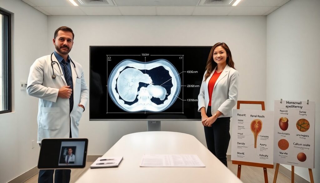
Imaging Modalities: CT, Ultrasound, X-Ray, And When To Use Each
Non-contrast CT serves as the primary test for suspected pielolitiase. CT shows stone size, density, and obstruction. Ultrasound suits pregnant patients and children. Ultrasound detects hydronephrosis and many pelvic stones but misses small stones. Plain abdominal X-ray (KUB) detects radiopaque stones such as calcium stones. Clinicians use CT when they need high sensitivity and to plan intervention. Ultrasound and KUB serve for follow-up and lower-risk scenarios. Low-dose CT protocols reduce radiation while maintaining accuracy.
Laboratory Tests And Stone Analysis: What Clinicians Look For
Clinicians order serum creatinine, electrolytes, and complete blood count. They obtain urinalysis and urine culture to detect infection. A positive culture with stones requires prompt drainage and antibiotics. Providers collect 24-hour urine when the patient recovers to measure volume, calcium, oxalate, citrate, sodium, and uric acid. Stone composition analysis informs prevention plans. For example, high urinary calcium leads to thiazide consideration. Low citrate suggests citrate supplementation. The lab data links cause to tailored prevention.
Treatment And Prevention Strategies: From Watchful Waiting To Surgery
Treatment for pielolitiase depends on size, location, symptoms, and infection. Small stones under 5 mm often pass with fluids and pain control. Alpha-blockers may speed passage for stones in the ureter. Stones 5–20 mm may need extracorporeal shock wave lithotripsy (ESWL) or ureteroscopy. Large or complex pelvic stones may require percutaneous nephrolithotomy (PCNL). Infection with obstruction needs urgent drainage with stent or nephrostomy and antibiotics. Prevention focuses on increased fluid intake, dietary changes, and medical therapy based on stone type. Patients who form calcium stones may reduce sodium and maintain normal calcium intake. Uric acid stones respond to urine alkalinization and xanthine oxidase inhibitors when indicated. Regular follow-up imaging and metabolic testing reduce recurrence. Clinicians personalize prevention using stone analysis and 24-hour urine results.